Home / Articles / Retinopathy of Prematurity (ROP)


Dr Süleyman Karaatlı and Dr Cüneyt Özmen
performing laser theraphy for ROP
During development, blood vessels grow from the central part of the retina outwards. This process is completed a few weeks before the normal time of delivery. However, inpremature babies it is incomplete. If blood vessels grow normally, ROP does not occur. If the vessels grow and branch abnormally the baby develops ROP. These abnormal blood vessels may grow up from the plane of the retina and may bleed inside the eye. When the blood and abnormal vessels are reabsorbed, it may give rise to multiple band likemembranes which can pull up the retina, causing detachment of the retina and eventually blindness before 6 months.
Normally, maturation of the retina proceeds in-utero, and at term, the medial portion of the retina is fully vascularized, while the lateral portion is only incompletely vascularized. The normal growth of the blood vessels is directed to relatively low-oxygen areas of the retina, but the vessels remain in the plane of the retina and do not grow into the vitreous humor. If excess oxygen is given, normal blood vessels degrade and cease to develop. When the excess oxygen environment is removed, the blood vessels rapidly begin forming again and grow into the vitreous humor of the eye from the retina.
The key disease element in ROP is fibrovascular proliferation. This is growth of abnormal new vessels; this may regress, but frequently progresses. Associated with the growth of these new vessels is fibrous tissue (scar tissue) that may contract to cause retinal detachment. Multiple factors can determine whether the disease progresses, including overall health, birth weight, the stage of ROP at initial diagnosis, and the presence or absence of "plus disease". Supplemental oxygen exposure, while a risk factor, is not the main risk factor for development of this disease. Restricting supplemental oxygen use reduces the rate of ROP, but may raise the risk of other hypoxia-related systemic complications, including death.
Patients with ROP, particularly those who have developed severe disease needing treatment are at greater risk for strabismus, glaucoma, cataracts and shortsightedness (myopia) later in life and should be examined yearly to help prevent or detect and treat these conditions.
Almost all infants with ROP have a gestational age of 31 weeks or less (regardless of birth weight) or a birth weight of 1250 g or less; these indications are generally used to decide whether a baby should be screened for ROP, but some centres, especially in developing countries extend birth weight screening criteria to 1500 gr.
Any premature baby with severe illness in perinatal period (Respiratory distress syndrome, sepsis, blood transfusion, Intra ventricular haemorrhage, apnoeic episodes, etc.) may also be offered ROP screening.
Retinal examination with scleral depression is generally recommended for patients born before 30–32 weeks gestation, or 4–6 weeks of life, whichever is later. It is then repeated every 1–3 weeks until vascularization is complete (or until disease progression mandates treatment).
Following pupillary dilation using eye drops, the retina is examined using a special lighted instrument (an indirect ophthalmoscope). The peripheral portions of the retina are sometimes pushed into view using scleral depression. Examination of the retina of a premature infant is performed to determine how far the retinal blood vessels have grown (the zone), and whether or not the vessels are growing flat along the wall of the eye (the stage). Once the vessels have grown into Zone 3 (see below) it is usually safe to discharge the child from further screening for ROP. The stage of ROP refers to the character of the leading edge of growing retinal blood vessels (at the vascular-avascular border).
The stages of ROP disease have been defined by the International Classification of Retinopathy of Prematurity (ICROP).
In older patients the appearance of the disease is less well described but includes the residua of the ICROP stages as well as secondary retinal responses.
The system used for describing the findings of active ROP is entitled The International Classification of Retinopathy of Prematurity (ICROP). ICROP uses a number of parameters to describe the disease. They are location of the disease into zones (1, 2, and 3), the circumferential extent of the disease based on the clock hours (1-12), the severity of the disease (stage 1-5) and the presence or absence of "Plus Disease". Each aspect of the classification has a technical definition. This classification was used for the major clinical trials. It was revised in 2005.

The zones are centered on the optic nerve. Zone 1 is the posterior zone of the retina, defined as the circle with a radius extending from the optic nerve to double the distance to the macula. Zone 2 is an annulus with the inner border defined by zone 1 and the outer border defined by the radius defined as the distance from the optic nerve to the nasal ora serrata. Zone 3 is the residual temporal crescent of the retina.
The circumferential extent of the disease is described in segments as if the top of the eye were 12 on the face of a clock. For example one might report that there is stage 1 disease for 3 clock hours from 4 to 7 o'clock. (The extent is a bit less important since the treatment indications from the Early Treatment for ROP)
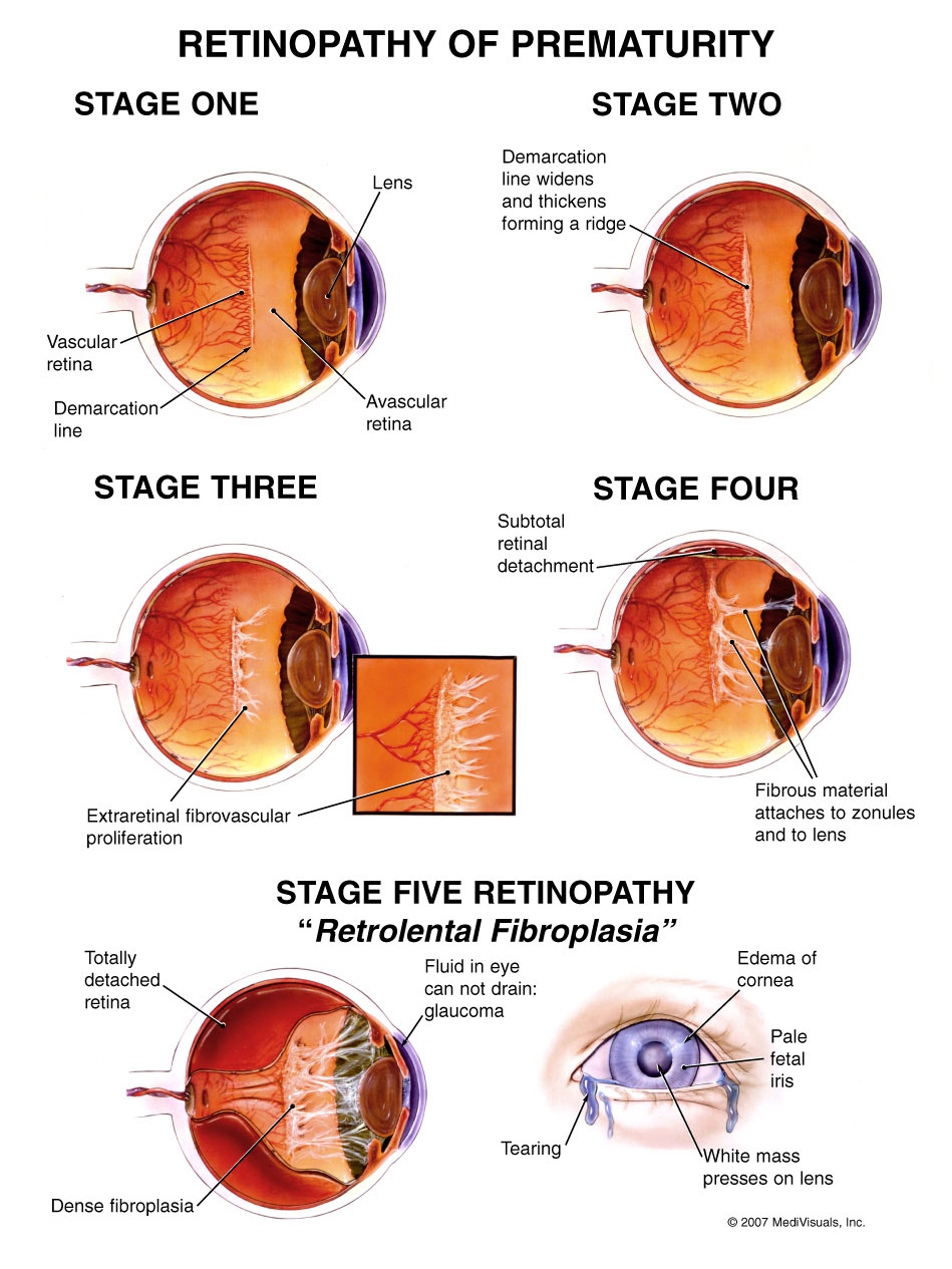
The Stages describe the ophthalmoscopic findings at the junction between the vascularized and avascular retina.
Plus disease can be present as a major complicating factor at any stage. It is characterised by:
Stages 1 and 2 do not lead to blindness. However, they can progress to the more severe stages. Threshold disease is defined as disease that has a 50% likelihood of progressing to retinal detachment. Threshold disease is considered to be present when stage 3 ROP is present in either zone I or zone II, with at least 5 continuous or 8 total clock hours of disease, and the presence of plus disease. Progression to stage 4 (partial retinal detachment), or to stage 5 (total retinal detachment), will result in substantial or total loss of vision for the infant.
The most difficult aspect of the differential diagnosis may arise from the similarity of two other diseases:
In order to allow timely intervention, a system of monitoring is undertaken for infants at risk of developing ROP. These monitoring protocols differ geographically because the definition of high-risk is not uniform or perfectly defined. In the USA the consensus statement of experts is informed by data derived by clinical trials and published in Pediatrics 2006. They included infants with birthweights under 1500 grams or under 30 weeks gestation in most cases. The first examination should take place within the first 4 weeks of life, and regular, weekly examination is required until it is clear that the eyes are not going to develop disease needing treatment, or one or both eyes develop disease requiring treatment. Treatment should be administered within a 48 hours, as the condition can progress rapidly.


This disease was first described in the premature baby in 1942. Between 1941–1953, over 12,000 babies worldwide were affected by it. Soul musician Stevie Wonder, actor Tom Sullivan as well as jazz singer Diane Schuur are a few famous people who have the disease.
The first case of the epidemic was seen on St. Valentine's Day in 1941, when a premature baby in Boston was diagnosed. Cases were then seen all over the world and the cause was, at that point, unknown. By 1951 a clear link between incidence and affluence became clear: many cases were seen in developed countries with organized and well-funded health care. Two British scientists suggested that it was oxygen toxicity that caused the disease. Babies born prematurely in such affluent areas were treated in incubators which had artificially high levels of oxygen. Studies on rats made this cause seem more likely, but the link was eventually confirmed by a controversial study undertaken by American pediatricians. The study involved two groups of babies. Some given the usual oxygen concentrations in their incubators, while the other group had "curtailed" oxygen levels. The latter group was shown to have a lower incidence of the disease. As a result, oxygen levels in incubators were lowered and consequently the epidemic was halted. Each case of ROP avoided by withholding oxygen "may have cost some 16 deaths".
ROP prevalence varies, from 5–8% in developed countries with adequate neonatological facilities, to up to 30% in middle-income developing countries.
There is increasing evidence that ROP and blindness due to ROP are now public health problems in the middle income countries of Latin America, Eastern Europe and the more advanced economies in South East Asia and the Middle east region. In these countries ROP is often the most common cause of blindness in children. ROP is highly likely to become an increasing problem in India, China and other countries in Asia as these countries expand the provision of services for premature infants.
There is also evidence that the population of premature infants at risk of severe ROP varies depending on the level of neonatal intensive care being provided. In countries with high development indices and very low neonatal mortality rates (e.g. North America, western Europe), severe ROP is generally limited to extremely preterm infants i.e. those weighing less than 1000g at birth. At the other end of the development spectrum, countries with very low development indices and very high neonatal mortality rates (e.g. much of subSaharan Africa) ROP is rare as most premature babies do not have access to neonatal intensive care and so do not survive. Countries with moderate development indices are improving access to neonatal intensive care, and in these settings bigger, more mature babies are also at risk of severe ROP as neonatal care may be suboptimal(i.e. those weighing 1500–2000 g at birth). These findings have two main implications: firstly, much can be done in countries with moderate development indices to improve neonatal care, to reduce the risk of severe ROP in bigger babies and increase survival of extremely preterm infants, and secondly, in these settings bigger more mature babies need to be included in ROP programs and examined regularly so as to detect those babies developing ROP requiring treatment.
In 2012, the World Health Organization published data on rates of preterm birth and the number of premature babies born in different regions of the world. The main findings of this report are threefold: 1. premature birth has many different causes, and prevention is challenging 2. prematurity is the commonest cause of neonatal death in many countries: 1 million infants die every year due to complications of preterm birth, and 3. the number of preterm births is currently estimated to be 15 million, and increasing.